IMR Press / JIN / Volume 23 / Issue 5 / DOI: 10.31083/j.jin2305091
Open Access Review
The Role of Microglia with Mitochondrial Dysfunction and Its Therapeutic Prospects in Alzheimer's Disease
Show Less
1 Department of Clinical Laboratory, Shenzhen Traditional Chinese Medicine Hospital, 518033 Shenzhen, Guangdong, China
2 Shenzhen Key Laboratory of Drug Addiction, Shenzhen Neher Neural Plasticity Laboratory, The Brain Cognition and Brain Disease Institute, Shenzhen Institute of Advanced Technology, Chinese Academy of Sciences, 518055 Shenzhen, Guangdong, China
*Correspondence: yl.xu1@siat.ac.cn (Yunlong Xu); zxh419@163.com (Xuehui Zeng); fuxiang2004@126.com (Fuxiang Zheng)
J. Integr. Neurosci. 2024, 23(5), 91; https://doi.org/10.31083/j.jin2305091
Submitted: 27 November 2023 | Revised: 28 January 2024 | Accepted: 26 February 2024 | Published: 30 April 2024
Copyright: © 2024 The Author(s). Published by IMR Press.
This is an open access article under the CC BY 4.0 license.
Abstract

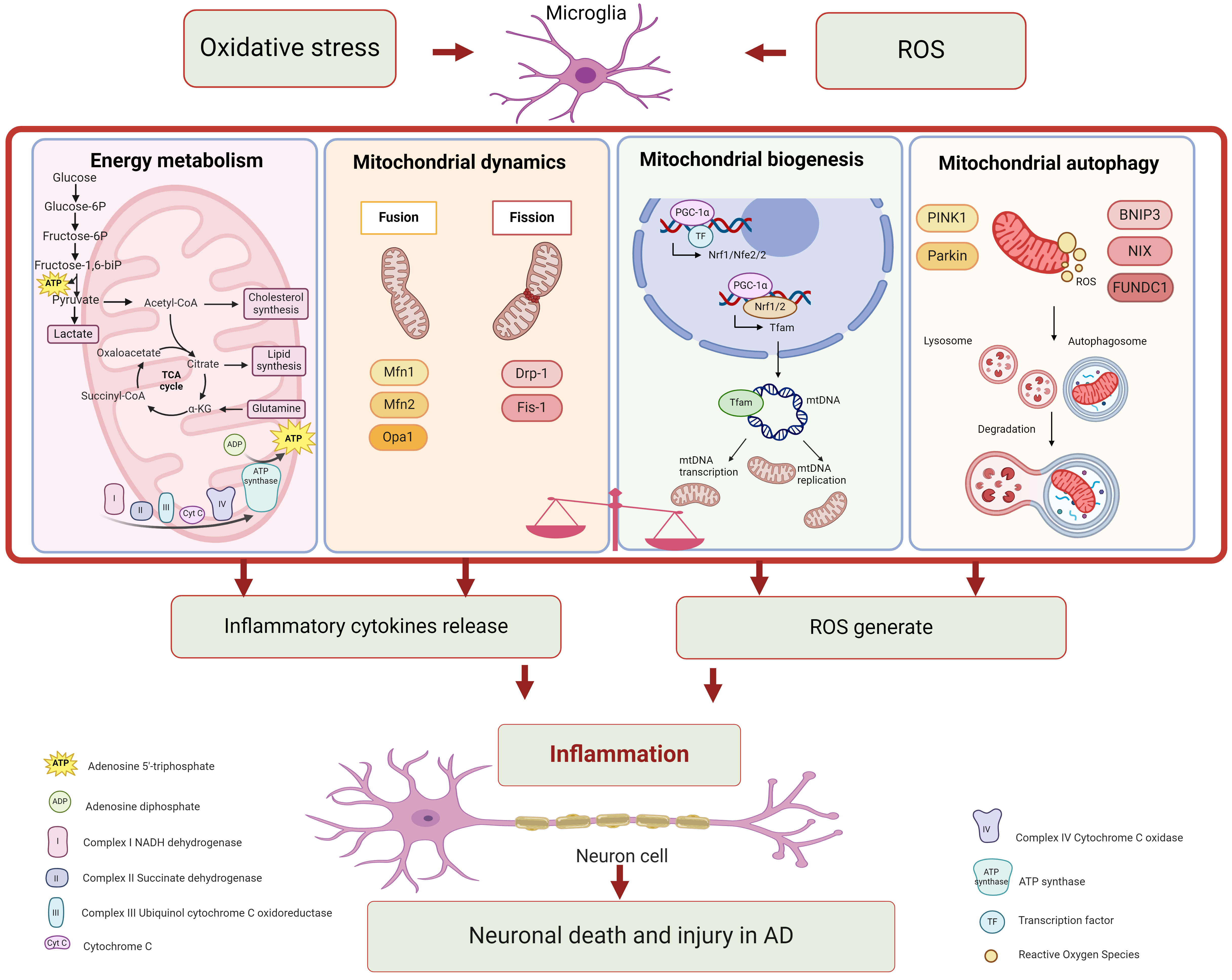
Alzheimer’s disease (AD), a primary cause of dementia, is rapidly emerging as one of the most financially taxing, lethal, and burdensome diseases of the 21st century. Increasing evidence suggests that microglia-mediated neuroinflammation plays a key role in both the initiation and progression of AD. Recently, emerging evidence has demonstrated mitochondrial dysfunction, particular in microglia where precedes neuroinflammation in AD. Multiple signaling pathways are implicated in this process and pharmaceutical interventions are potentially involved in AD treatment. In this review, advance over the last five years in the signaling pathways and pharmaceutical interventions are summarized and it is proposed that targeting the signaling pathways in microglia with mitochondrial dysfunction could represent a novel direction for AD treatment.

Keywords
Alzheimer's disease
microglia
mitochondria
microglia with mitochondrial dysfunction
Funding
JCYJ20210324111213037/Shenzhen Science and Technology Program
2023M733658/China Postdoctoral Science Foundation
Figures
Fig. 1.
Share
Back to top